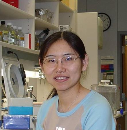
佟超

佟超 博士
浙江大学生命科学研究院教授、研究员、博士生导师
办公地点:医学院科研楼A401
电话: 0571-88981582 (office)
传真: 0571-88981582
Email: ctong@zju.edu.cn
实验室网站:http://lsi.zju.edu.cn/yjdw_detail.aspx?ID=16
详细介绍:
教育和工作背景
1995-1999:南开大学 生化及分子生物学系 学士
1999-2002: 中国科学院动物研究所 生殖生物学 硕士
2002-2006: 美国得克萨斯大学西南医学中心(UTSW)遗传及发育生物学 博士
2007-2011: 美国贝勒医学院( Baylor College of Medicine) 人类分子遗传学系 博士后
2011-至今: 浙江大学生命科学研究院教授,研究员,博士生导师
学术奖项与活动
2000:中国科学院董事东方奖学金
2002:中国科学院院长奖学金优秀奖
2007:Developmental biology training grant
2008: “Brain disorders and Development” training grant
2004-至今:member of Genetics society of America
研究方向
我们研究组致力于研究神经系统发育及神经退行性疾病的分子机制。我们主要感兴趣的问题是神经细胞轴突如何找到其靶细胞并与之建立精确突触连接,以及细胞自
噬通路如何维持神经细胞完整性防止神经退行性疾病的发生。我们的目标是通过果蝇遗传筛选的方法寻找参与神经轴突定位及神经细胞完整性维持的新基因,阐明其
作用的分子机理,
并最终在高等动物中检验此作用机制的保守性。从而为临床实践提供理论依据。同时,我们也希望我们的研究能够拓展人们对细胞之间相互作用以及细胞自噬等基本
细胞生物学过程的了解,为攻克人类疾病做出贡献。
一、神经元轴突定位
在大脑发育过程中,10亿个神经元整合多种信号相互连接形成约1兆个神经突触。神经元轴突寻找其靶细胞并与之建立精确突触连接对于神经系统的发育和神经回
路的建立至关重要。而人们对于神经元如何整合外界信号从而精密调节其轴突行为的了解还非常有限。近年研究表明,不正常的轴突定向及突触连接可以导致多种神
经系统先天疾病,例如智障,自闭症,以及神经退行性疾病。因此,研究神经元轴突定向及突触连接建立的分子机理仍是当今神经生物学的重要任务之一。果蝇的视
觉神经系统因其结构的模块性,进化上的保守性,以及实验上的可操作性为我们解析神经元轴突定向及突触连接建立的分子机理提供了一个极好的模型。我们在前期
工作中,通过大规模遗传筛选发现一个进化上保守的新基因rich调节感光神经细胞R7的轴突定向。Rich蛋白与Rab6结合并正向调节Rab6的活性,
进而影响细胞表面粘联蛋白N-Cadherin 的分布,造成R7轴突定位异常。我们将进一步研究N-Cadherin
在细胞中的运输方式,继续研究Rich 如何调节Rab6活性, 并进而研究Rich 哺乳动物同源基因的功能。
二、细胞自噬与神经退行性疾病
随着人类寿命的增长,神经退行性疾病的发生日益普遍,严重影响人类生活质量。然而,人们对神经退行性疾病发生发展的分子机理还知之甚少。神经元细胞寿命很
长,又不能通过细胞分裂稀释细胞中异常的蛋白和损坏的细胞器。因此识别并清除这些废物,防止他们的堆积造成的细胞毒性对神经细胞来讲十分重要。近年研究表
明,细胞自噬功能异常在神经退行性疾病中相当普遍。并且,在小鼠和果蝇中敲除细胞自噬相关基因,均可诱导神经退行性疾病类似症状。重要的是,人们发现调节
细胞自噬功能可以减缓多种神经退行性疾病症状。因此研究细胞自噬这一生物过程,寻找新的调节细胞自噬的基因,对我们阐明神经退行性疾病的分子机理,探索新
的治疗方案都至关重要。
我们在前期工作中通过大规模遗传筛选,发现多个果蝇突变株具有细胞自噬障碍。其中一些基因的突变在人类中导致神经退行性疾病,但其功能从未与细胞自噬连系
起来。对这些基因的研究将为我们阐明这些神经退行性疾病的致病机理提出治疗方案提供帮助。此外,我们还发现同是编码线粒体蛋白的基因,
有些基因的突变会促进细胞自噬,而另外一些的突变却抑制细胞自噬。
因为线粒体是通过细胞自噬进行清除的,对这些基因的进一步的研究将有助于我们了解细胞自噬的信号传导和底物识别将有很大帮助。此外在我们已经筛出的基因中
还有参与细胞中重要信号通路的分子,参与细胞内膜泡运输的分子,以及未知功能的新基因。进一步研究这些分子在细胞自噬中的作用将为我们全面了解这一重要生
物过程开启新的一页。
Research interests:
Our major research interest is to dissect the molecular mechanisms that
underline neuronal development and neural degeneration diseases.
Particularly, I’m interested in how neurons find their targets and how
autophagy/lysosomal pathway contribute to neuronal health maintenance.
Our goal is to identify novel players and to define molecular pathways
in neural development and maintenance by using Drosophila as a model
system. Eventually, we want to apply these discoveries to the higher
organisms. We hope our study can provide clues for clinical treatments
of cognitive diseases and neural degeneration. Meanwhile, we hope our
study could expand our understanding of the general autophagy/lysosomal
pathway which is also involved in multiple important physiological and
pathological processes such as immune-response and cancer in addition to
neural health maintenance.
Neuronal specificity
During development, more than 100 billon neurons in our brain
form trillions synaptic connections. To achieve this, neurons integrate
numerous signals that allow them to decide when to extend their growth
cones, to follow a specific route, to determine when to fasciculate or
defasciculate, and when to stop and form synaptic connections. Improper
synapse formation may lead to cognitive diseases and mental retardation
such as autism spectrum disorders. However, we still know very little
about how neurons form such complex network. Hence, providing a
molecular framework to understand how neurons form proper synapses with
their targets remains an important endeavor.
The Drosophila visual system is an excellent model to untangle this type
of question. By using forward genetic screen, we identified mutations
in rich, a novel gene that is evolutionarily conserved from worms to
human. We found that rich is required for synaptic specificity in
Drosophila eyes and olfactory neurons and regulate Rab6 activity. Our
data define a novel role for Rich and Rab6 in regulating synaptic
specificity by regulating trafficking of CadN but not other proteins
implicated in this process.
In the future, we’ll further investigate how CadN is trafficked inside
cells. We’ll also like to further understand the molecular nature of
Rich and elucidate how Rich regulates Rab6. In addition, we also like to
study the functions of the mammalian homolog.
Neuodegeneration and autophagy/lysosomal pathway
At adulthood, neurons have to maintaining cell integrity in
response to genetic and environment insults to sustain nerve system
function and prevent degeneration. With increase of human life-span,
neural-degeneration emerged as rather common diseases that greatly
affect people’s life quality. However, the molecular mechanisms
underline the neurodegeneration diseases are poorly understood.
Drosophila has proved to be an excellent model to study
neurodegeneration.
As long-lived post-mitotic cells, neurons cannot dilute the altered
proteins and damaged organelles by cell division. Therefore, to
identify and reduce these malfunction structures before they buildup and
cause neurotoxicity is particular important to neurons.
Autophagy/lysosomal pathway plays important role in this clearance
process, evidenced by autophagic malfunction in many human neurological
disorders such as Alzheimer’s disease, Parkinson’s disease, Huntington’s
disease, and amyotrophic lateral sclerosis (ALS). In addition,
genetically deleted autophagy related genes (Atgs) in mice and flies
results neural degeneration. In contrast, modulating autophagy has
beneficial effects in many neurodegenerative disorders. Therefore,
understanding autophagy/lysosomal pathway should facilitate the
neurodegeneration study.
We carried out a forward genetic screen to identify novel player
involved in autophagy /lysosomal pathway. So far, we have identified
more than 60 mutant complementation groups with autophagy defects. Among
these mutants, some genes have already been implied involved in
neurodegeneration diseases in human. But their functions have never been
linked to autophagy pathway. Therefore, study the function of these
genes in autophagy pathway will be important for us to understand the
molecular mechanisms underline these diseases. We also find genes
encoding mitochondrial proteins play diverse roles in autophagy pathway,
some mutants block autophagy while the others enhance autophay. It will
be very interest to study how mitochondrial proteins regulate autophagy
pathway. In addition, we also found proteins involved in signal
transduction, membrane trafficking, as well as proteins with unknown
function. We believe with these studies we’ll expand our understanding
of autophagy pathway as well as neurodegeneration.
发表论文
1. Tong, C.,
Ohyama, T., Tie, A., Rajan, A., Haueter, C., Bellen, HJ. Rich regulates
target specificity of photoreceptor cells and N-Cadherin trafficking in
the Drosophila visual system via Rab6. Neuron Accepted
2. Jia, H., Liu, Y., Xia, R., Tong, C., Yue, T., Jiang, J., Jia, J. (2010). Casein kinase 2 promotes hedgehog signaling by regulating both smoothened and cubitus interruptus. J Biol Chem. 285(48):37218-26.
3. Chen, Y., Li, S., Tong, C., Zhao, Y., Wang, B., Liu, Y., Jia, J., Jiang, J. (2010). G protein-coupled receptor kinase 2 promotes high-level Hedgehog signaling by regulating the active state of Smo through kinasedependent and kinase-independent mechanisms in Drosophila. Genes Dev. 24(18):2054-67.
4. Bellen, HJ., Tong, C., Tsuda, H. (2010). 100 years of Drosophila research and its impact on vertebrate neuroscience: a history lesson for the future. Nat Rev Neurosci. 11(7), 514-522
5. Tsuda, H., Han, SM., Yang, Y., Tong, C., Lin, YQ., Mohan, K., Haueter, C., Zoghbi, A., Harati, Y., Kwan, J., Miller, MA., Bellen HJ. (2008). The amyotrophic lateral sclerosis 8 protein VAPB is cleaved, secreted, and acts as a ligand for Eph receptors. Cell. 133(6), 963-77.
6. Zhao, Y., Tong, C., Jiang, J. (2007). Transducing the Hedgehog signal across the plasma membrane. Fly.1(6), 333-6.
7. Tong, C., Jiang, J. (2007). Using immunoprecipitation to study protein-protein interactions in the hedgehogsignaling pathway. Methods Mol Biol. 397, 215-30.
8. Zhao, Y.*, Tong, C*., Jiang, J. (2007). Hedgehog regulates smoothened activity by inducing a conformational switch. Nature 450, 252-8. (*共同第一作者)
9. Jia, J., Zhang, L., Zhang, Q., Tong, C., Wang, B., Hou, F., Amanai, K., and Jiang, J. (2005).Phosphorylation by double-time/CKIepsilon and CKIalpha targets cubitus interruptus for Slimb/beta-TRCPmediated proteolytic processing. Developmental Cell 9, 819-830.
10. Zhang, W., Zhao, Y., Tong, C., Wang, G., Wang, B., Jia, J., and Jiang, J. (2005). Hedgehog-regulated Costal2-kinase complexes control phosphorylation and proteolytic processing of Cubitus interruptus. Developmental Cell 8, 267-278.
11. Jia, J.*, Tong, C.*, Wang, B., Luo, L., and Jiang, J. (2004). Hedgehog signalling activity of Smoothened requires phosphorylation by protein kinase A and casein kinase I. Nature 432, 1045-1050. (* 共同第一作者)
12. Jia, J., Tong, C., and Jiang, J. (2003). Smoothened transduces Hedgehog signal by physically interacting with Costal2/Fused complex through its C-terminal tail. Genes and Development 17, 2709-2720.
13. Moskalenko, S., Tong, C., Rosse, C., Mirey, G., Formstecher, E., Daviet, L., Camonis, J., and White, M. A. (2003). Ral GTPases regulate exocyst assembly through dual subunit interactions. The Journal of biological chemistry 278, 51743-51748.
14. Tong, C., Fan, H. Y., Chen D, Y., Song, X. F., Schatten, H., and Sun, Q. Y. (2003). Effects of MEK inhibitor U0126 on meiotic progression in mouse oocytes: microtuble organization, asymmetric division and metaphase II arrest. Cell Research 13, 375-383.
15. Fan, H. Y., Tong, C., Teng, C. B., Lian, L., Li, S. W., Yang, Z. M., Chen, D. Y., Schatten, H., and Sun, Q.Y. (2003). Characterization of Polo-like kinase-1 in rat oocytes and early embryos implies its functional roles in the regulation of meiotic maturation, fertilization, and cleavage. Molecular Reproduction and Development 65, 318-329.
16. Fan, H. Y., Tong, C., Lian, L., Li, S. W., Gao, W. X., Cheng, Y., Chen, D. Y., Schatten, H., and Sun, Q. Y. (2003). Characterization of ribosomal S6 protein kinase p90rsk during meiotic maturation and fertilization in pig oocytes: mitogen-activated protein kinase-associated activation and localization. Biology of Reproduction 68, 968-977.
17. Cheng, Y., Fan, H. Y., Wen, D. C., Tong, C., Zhu, Z. Y., Lei, L., Sun, Q. Y., and Chen, D. Y. (2003). Asynchronous cytoplast and karyoplast transplantation reveals that the cytoplasm determines the developmental fate of the nucleus in mouse oocytes. Molecular Reproduction and Development 65, 278-282.
18. Yao, L. J., Fan, H. Y., Tong, C., Chen, D. Y., Schatten, H., and Sun, Q. Y. (2003). Polo-like kinase-1 in porcine oocyte meiotic maturation, fertilization and early embryonic mitosis. Cellular and molecular biology (Noisy-le-Grand, France) 49, 399-405.
19. Fan, H. Y., Li, M. Y., Tong, C., Chen, D. Y., Xia, G. L., Song, X. F., Schatten, H., and Sun, Q. Y. (2002). Inhibitory effects of cAMP and protein kinase C on meiotic maturation and MAP kinase phosphorylation in porcine oocytes. Molecular Reproduction and Development 63, 480-487.
20. Fan, H. Y., Tong, C., Li, M. Y., Lian, L., Chen, D. Y., Schatten, H., and Sun, Q. Y. (2002). Translocation of the classic protein kinase C isoforms in porcine oocytes: implications of protein kinase C involvement in the regulation of nuclear activity and cortical granule exocytosis. Experimental Cell Research 277, 183-191.
21. Tong, C., Fan, H. Y., Lian, L., Li, S. W., Chen, D. Y., Schatten, H., and Sun, Q. Y. (2002). Polo-like kinase-1 is a pivotal regulator of microtubule assembly during mouse oocyte meiotic maturation, fertilization, and early embryonic mitosis. Biology of Reproduction 67, 546-554.
附件列表
词条内容仅供参考,如果您需要解决具体问题
(尤其在法律、医学等领域),建议您咨询相关领域专业人士。
